| Search for content and authors |
Up-conversion and down-conversion processes observed in Er3+, Yb3+ and Mn2+ doped ZnAl2O4 nanoparticles |
| Izabela Kamińska 1, Krzysztof Fronc 1, Bożena Sikora 1, Anna Baranowska-Korczyc 1, Kamil Sobczak 1, Tomasz Wojciechowski 1, Wojciech Paszkowicz 1, Roman Minikayev 1, Mateusz Chwastyk , Kamil Koper 2,3, Piotr Stępień 2,3, Bohdan Paterczyk 4, Grzegorz M. Wilczyński 5, Jakub Włodarczyk 5, Maciej Gawlak 5, Danek Elbaum 1 |
|
1. Polish Academy of Sciences, Institute of Physics, al. Lotników 32/46, Warszawa 02-668, Poland |
| Abstract |
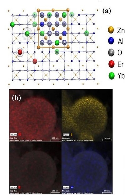
Our objective was to synthesize hydrophilic zinc-aluminum spinel nanostructures doped with rare earth ions such as Er3+ and Yb3+ (fig. 1a) and ions from transition metal group such as Mn2+. Spinels are isomorphic metal oxides AB2O4, where A and B are divalent and trivalent ions, respectively. The material shows a close-packed face centered cubic structure with Fd3m space group symmetry. They are applicable as useful luminescent bio-marker due to a significant improvement in the signal to background ratio and high resilience to photo-bleaching.
Fig. 1. (a) Spinel ZnAl2O4: Er3+, Yb3+ unit cell geometry (upconversion process) [2], (b) Chemical distribution maps of elements: Zn, O, Er, Yb. A wide variety of emission spectra could be obtained by changing the concentrations and proportions of the rare earth ions in the crystal host. Although upconversion can be expected, in principle, from most lanthanide-doped crystalline host materials, efficient UC occurs only by using a limited number of well selected host-dopant combinations [1]. The nanoparticles were synthesized in aerosol solutions consisting of droplets (4μm in diameter) by injecting a solution containing the reductors and oxidants to a furnace at the temperature of 1000oC. The structures of the materials were characterized by transmission electron microscopy and X-ray diffraction. We obtained regular spherical polycrystalline nanoparticles with a broad size distribution from 20 to 800 nm. To evaluate morphological changes of nanoparticles surface and to visualize the surface topography we used scanning electron microscope. In addition, we performed the EDX material analysis in order to determine distribution of elements in the samples. The results confirmed the presence of Zn, Al, O and Er and Yb (fig. 1b). To check the toxicity of the ZnAl2O4: Er3+, Yb3+ nanoparticles, we introduced them to the body of the living nematode (Caenorhabditiselegans). C. elegans is a frequently used model organism in biological research, such as process development, embryogenesis, morphogenesis and aging. Confocal microscope images confirm the presence of the nanoparticles inside the gastrointestinal tract of C. elegans.References [1] Wang F., Liu X., Chem. Soc. Rev. 2009, 38, 976-989. [2] Humphrey W., Dalke A., Schulten K.,VMD -Visual Molecular Dynamics, J. Molecular Graphics, 1996, 14, 33-38.
The research was supported by the European Union within European Regional Development Fund, through grant Innovative Economy (POIG.01.01.02-00-008/08). The Polish National Centre for Research and Development NR13004704 and Center of Excellence. This work was (partially) performed in the NanoFun laboratories co-financed by the European Regional Development Fund within the Innovation Economy Operational Programme, the Project No. POIG.02.02.00-00-025/09/.
|
| Legal notice |
|
| Related papers |
Presentation: Poster at Nano-Biotechnologia PL, by Izabela KamińskaSee On-line Journal of Nano-Biotechnologia PL Submitted: 2012-06-28 11:25 Revised: 2012-09-13 07:54 |