| Search for content and authors |
Synthesis and properties of NaYF4: Er, Yb, Gd nanoparticles with and without SiO2 coating for biomedical applications. |
| Bożena Sikora 1, Sebastian Szewczyk 2, Krzysztof Fronc 1, Izabela Kamińska 1, Anna Baranowska-Korczyc 1, Kamil Koper 3,4, Kamil Sobczak 1, Jakub Włodarczyk 5, Roman Minikayev 1, Wojciech Paszkowicz 1, Tomasz Wojciechowski 1, Grzegorz M. Wilczyński 5, Piotr Stępień 3,4, Danek Elbaum 1 |
|
1. Polish Academy of Sciences, Institute of Physics, Lotnikow 32/46, Warsaw 02-668, Poland |
| Abstract |
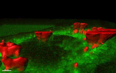
Cell autofluorescence is an important limitation in majority of applications based on luminescent probes. In addition, most biological structures absorb ultraviolet and visible light. On the other hand, significantly lower absorption and resulted autofluorescence of the near infrared energy (wavelength 700 - 1000 nm) is observed for biological materials. Our aim was to synthesize NaYF4 nanoparticles doped with up-converting rare earth metals(Er, Yb). The conversion takes place by a multiphoton absorption of a low-energy radiation (near infrared – optimum 980 nm) and subsequent emission of higher energy radiation (visible light). Synthesized up-converting nanoparticles were subsequently coated by a layer of SiO2 for the protection of biological systems from the toxic effects of fluoride and subsequent attachment to biologically active molecules. We synthesized NaYF4 nanoparticles doped by 2 % Er and various Yb concentrations (10 to 30%). Nanoparticles excited by infrared light (965 nm) have two luminescence bands: the first with a maximum at 540 nm and the second, more intense band, with a maximum at a wavelength of 670 nm. The most efficient luminescence was observed for nanoparticles doped with 2% Er and 30% Yb. Synthesized by us nanoparticles have a diameter from 35 to 60 nm as determined by a transmission electron microscopy (TEM) and X-ray diffraction. These methods have confirmed the cubic structure of the nanoparticles. Using TEM we were able to confirm the presence of a thin SiO2 layer on the surface of nanoparticles. The upconverting SiO2 coated nanoparticles were transported into HeLa cells and their luminescence was measured by a confocal microscopy (Figure 1). Subsequently, these nanoparticles were linked to biologically active molecules to created a biosensor based on the Fluorescent Resonant Energy Transfer between the nanoparticles (donors) and organic probe sensitive to the external environment (acceptors).
Figure 1. NaYF4: Er, Yb nanoparticles inside HeLa cells. Image obtained from confocal microscopy. Green color: autofluorescence of cells (excitation: 488 nm); red color: NaYF4 nanoparticles (excitation: 960 nm). Acknowledgements: The research was partially supported by the European Union within European Regional Development Fund, through grant Innovative Economy (POIG.01.01.02-00-008/08) and was partially supported by the Ministry of Science and Higher Education (Poland) through Grant No. N N518 424036, and grant from the Polish National Centre for Research and Development NR13004704. |
| Legal notice |
|
| Related papers |
Presentation: Poster at SMCBS'2011 International Workshop, by Bożena SikoraSee On-line Journal of SMCBS'2011 International Workshop Submitted: 2011-08-31 17:15 Revised: 2011-08-31 18:17 |