| Search for content and authors |
Structural, morphological and optical properties of ZnAl2O4 nanoparticles co-doped with Er3+and Yb3+ prepared by combustion aerosol synthesis. |
| Izabela Kamińska 1, Krzysztof Fronc 1, Bożena Sikora 1, Anna Baranowska-Korczyc 1, Kamil Sobczak 1, Anna Reszka 1, Wojciech Paszkowicz 1, Kamil Koper 2,3, Grzegorz M. Wilczyński 4, Jakub Włodarczyk 4, Andrzej Szczepankiewicz 4, Piotr Stępień 2,3, B. J. Kowalski 1, Danek Elbaum 1 |
|
1. Institute of Physics, Polish Academy of Sciences, Warsaw 02-668, Poland |
| Abstract |
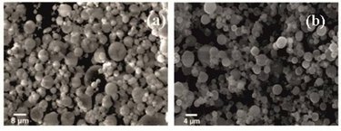
Zinc aluminate (ZnAl2O4) is found in nature as a mineral named gahnite. This spinel presents a close-packed face-centered cubic structure belonging to the Fd3m space group, having an optical band gap of 3.8 eV, which makes it suitable for photoelectronic devices, such as plasma display panels (PDPs), and field emission displays (FEDs). In recent years, several studies have been based on ZnAl2O4 doped rare earth ions (for example: Er3+, Yb3+, Dy3+, Tb3+, Eu3+, Tm3+). ZnAl2O4 co-doped Er3+and Yb3+ were prepared by a combustion aerosol process. The urea and nitrate compounds dissolved in distilled water and used for the production of aerosol were injected to a high– temperature reaction zone. As results we obtained spherical nanoparticles that up-converted infrared light (980nm) to visible light (540 nm and 650 nm). The synthesized nanopowders were characterized by: TEM, SEM, XRD, PL and confocal microscopy. TEM analysis showed a broad particle size distribution for (100 - 800 nm), for ZnAl1,9Er0,017Yb0,083O4 (Er:Yb = 1:5) for the oven calcined at 990oC for 3h in the air and unheated nanoparticles . Spherical polycrystalline nanoparticles were presented on the SEM photographs (Figure 1). In order to label cellular biological structures, the up-converting nanoparticles were transported into living HeLa cells and were detected by confocal microscopy. Our results indicate that the spinels can be visualized in HeLa cells. Fig. 1. SEM photographs of ZnAl2O4 co-doped Er3+and Yb3+ nanoparticles (a) calcined at 990 oC for 3h in air (b) not heated.
The research was supported by the European Union within European Regional Development Fund, through grant Innovative Economy (POIG.01.01.02-00-008/08), and by the Ministry of Science and Higher Education (Poland) through Grant No. N518 424036 and Center of Excellance. The Polish National Centre for Research and Development NR13004704 and Center of Excellence |
| Legal notice |
|
| Related papers |
Presentation: Poster at SMCBS'2011 International Workshop, by Izabela KamińskaSee On-line Journal of SMCBS'2011 International Workshop Submitted: 2011-08-31 16:06 Revised: 2011-09-09 11:45 |